by Laurence Garey and Eric Mensah-Brown
Department of Anatomy
Faculty of Medicine and Health Sciences
UAE University
PO Box 17666
Al Ain, United Arab Emirates
Summary
As part of the camel brain project supported by the Al Ain Chapter of ENHG,
we have studied the superior colliculus, a region on the surface of the
brainstem of the camel, Camelus dromedarius, using modern immunohistochemical
techniques. We had observed that, unlike in most mammals, the superior
colliculus of the camel is much larger than the inferior colliculus, another
similar nervous centre lying alongside it. It is known that the superior
colliculus is concerned with visual reflexes and head, neck and eye movements,
of obvious importance to this animal with large eyes, head and neck, and
apparent good vision. We looked at the distribution of neuropeptides, chemicals
that help the transmission of information around the brain. We describe for the
first time an unusually large content of neurons in the superior colliculus that
contain enkephalin, an opioid (morphine-like substance). We propose that this
system is associated with a pain-inhibiting pathway in the brain and spinal cord
that could be important in relation to the camel's life in the harsh environment
of its native deserts.
Introduction
The mammalian brain consists essentially of the cerebral hemispheres, covered
by the cerebral cortex, the cerebellum related largely to motor integration and
learning, and the brainstem which links the hemispheres to the spinal cord.
Whereas the hemispheres and cortex are the prime location for sensory
integration and motor activity, together with higher cognitive function, the
brainstem (medulla, pons and midbrain) contains neural centres that support
reflex activity. The superior and inferior colliculi form laminated eminences on
the dorsal surface of the midbrain. The superior colliculi are related to visual
reflexes, whereas the inferior colliculi form part of the auditory pathway.
Many studies have established the architecture of the colliculi in species
from rodents to man. However a perusal of the literature reveals a paucity of
information on large mammals, such as the camel. The camel is held in local
folklore to possess exceptional visual capabilities, a quality that has aided
its adaptation to the arid environment of the desert (Figure
1).
Results
We obtained the brains of adult male camels aged between 2 and 4 years from a
commercial abattoir. After standard laboratory preparation, we examined sections
of the colliculus with a microscope. We also made observations on skulls collect
from the local desert, and a brain kindly made available by Drs U Wernery and J
Kinne.
The cranial cavity of the camel is small compared with the whole skull (Figure 2). Its brain is also relatively small, up to
about 500g (Figure 3), considering its large total
body mass. We were surprised to find that its superior colliculus was several
times the volume of the inferior colliculus (Figure 3), whereas as in most
species (including man) the two are approximately the same size (Figure 4). . This difference in size could be
significant because the dolphin, an ungulate like the camel, is known to have an
elaborate auditory system and possesses an inferior colliculus that is much
larger than the superior colliculus (Figure 5). So,
the main aim of our study was to determine to what extent the very large
superior colliculus of the camel might have a special neuronal architecture or
distribution of neurotransmitters that are responsible for passage of neural
information between nerve cells.
The superior colliculus is composed of layers of alternating grey matter
(largely composed of neuronal cell bodies) and white matter (containing mainly
nerve fibres) (Figure 6). The superficial layers
receive nerve fibres (axons) from the retina of the eye (Figure
7) and from the visual cortex, the highest processing area for visual
information coming from the retina. The superior colliculus is also involved in
processing sensory information from other modalities than just visual, including
feedback to its deeper layers from the auditory, general sensory and motor
regions of the cerebral cortex. This is consistent with the superior colliculus
being an important centre for the control of orientation of head and eye.
We found that the neuronal architecture of the superior colliculus of the
camel is similar to that of other mammals. Neuronal cell bodies containing
certain common neuropeptides are limited to the superficial layers (Figure 8), whereas others are restricted to deeper
layers, and yet others are only in the grey matter deep to the superior
colliculus, the so-called periaqueductal grey (PAG).
Of the peptides we studied, the most interesting were the enkephalins (or
endorphins). These "intrinsic opioids" are important in the regulation
of pain. In particular, met-enkephalin neurons showed striking differences from
what has been described before. Most were small, and predominantly in the
superficial layers, but some larger met-enkephalin neurons were observed,
especially in the intermediate grey layer ( (Figure 9), (Figure 10)). Long enkephalin fibres were observed
bridging between the superficial, intermediate and deep layers, while some
extended into the PAG (Figure 11).
Discussion
The very large size of the superior colliculus of the camel brain suggests it
may have a special function in this animal. This may be related to a
well-developed visual system, and the necessity for the camel to coordinate eye
and head and movements accentuated by its long, muscular neck. However, our
discovery of numerous large met-enkephalin neurons in the superior colliculus
suggests a specialisation of this part of the brain compared with other mammals
studied so far.
Previously, enkephalins in the superior colliculus have been described mainly
in a minor population of small neurons in the superficial layers. We confirm
this presence of small enkephalin neurons in the superficial, visual layers.
However, in the camel a considerable population of large enkephalin neurons is
found in the deep layers. Enkephalins are important in pain suppression in
central neural pathways: they are endogenous opiates. They may explain the
well-known phenomenon of initial inhibition of pain sensation that can last for
several hours after an injury. A pain inhibitory pathway (Figure
12) has been described from the PAG to the lower brainstem, and thence to
the spinal cord. We propose that the presence of numerous large enkephalin
neurons throughout the superior colliculus with fibres projecting to the PAG
suggests that the pain inhibiting opioid pathway may be especially well
developed in the camel, perhaps helping to suit it for the extremes of
temperature and discomfort one might expect it to suffer in the desert.
Acknowledgements
This work was supported by grants from the UAE University and the Emirates
Natural History Group, Al Ain. We thank Ulrich Wernery and Joerg Kinne for help
with initial pilot experiments. Our special thanks are due to the late Alyaa Ali
Hussain Talib without whose devotion these observations would never have been
made.
Figures
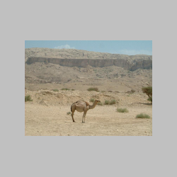
Figure 1. The camel in typical desert surroundings. There is little
protection from the sun at any time of the day. |
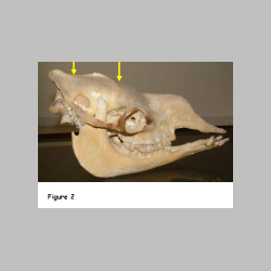
Figure 2. The skull of the camel has rather little space for the brain
(between the arrows). |
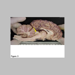
Figure 3. The camel brain is like most ungulate brains and only weighs
about 500g in spite of the large body mass. The very large superior colliculus
(arrow) is obvious (specimen with grateful thanks to U Wernery and J Kinne). |
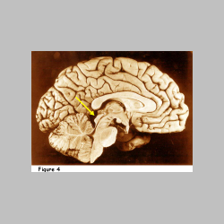
Figure 4. Medial view of the human brain. Note that the colliculi
(arrowed) are about the same size. |

Figure 5. In the dolphin, the inferior colliculus (IC) is much larger than
the superior (SC). This may be related to the important auditory functions of
this animal (eg. sonar). |
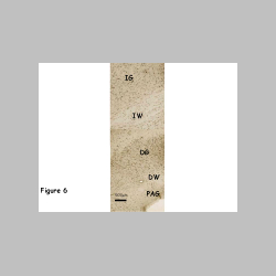
Figure 6. An autoradiograph of the superior colliculi of a monkey in which
a radioactive marker had been injected in the retina. The marker (glowing
brightly in this "dark field" micrograph) has been transported along
the axons from the retina to the superficial laminae of the colliculus. |
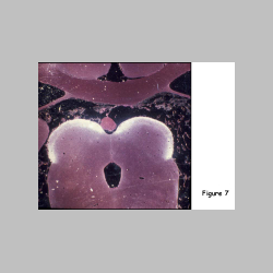
Figure 7. Low power micrograph of the intermediate grey (IG) and white
(IW), and deep grey (DG) and white (DW) laminae of the superior colliculus, plus
the periaqueductal grey (PAG). |
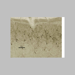
Figure 8. Specific neuropeptides are found mainly in restricted layers, in
this case substance P neurons in the superficial grey. |
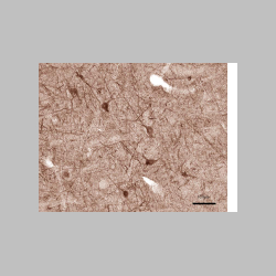 |
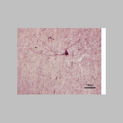
Figures 9,10. Large met-enkephalin neurons in the intermediate grey. |
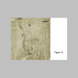
Figure 11. Bridging met-enkephalin axons between the intermediate grey and
PAG. |
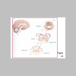
Figure 12. Schema of the pain inhibitory pathway (from Bear MF, Connors
BW, Paradiso MA, 2001. Neuroscience. Exploring the Brain. Second edition.
Baltimore: Lippincott, Williams and Wilkins) |